What is Osteoporosis?
A silent disease where the skeleton's internal architecture erodes until ordinary loads break bone.
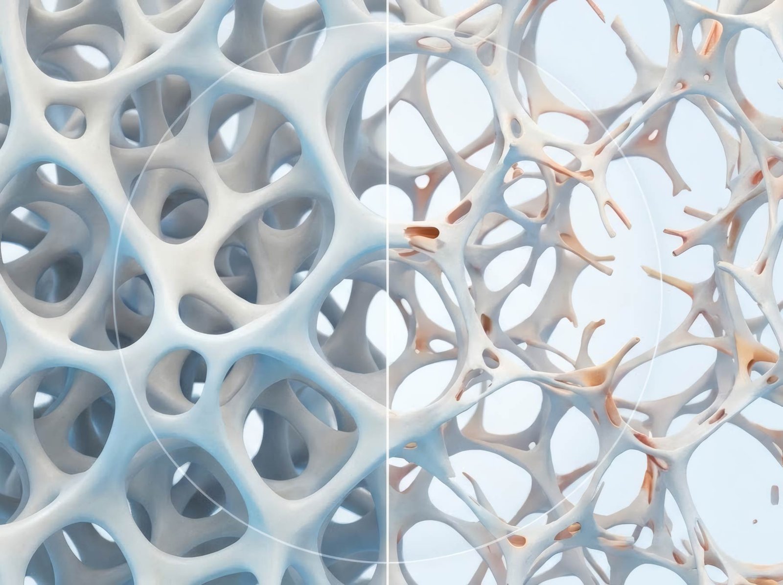
Osteoporosis — literally 'porous bone' — is a skeletal disease in which bone mineral density and micro-architecture deteriorate until a fall from standing height, a hug, or a cough can fracture a bone. It affects an estimated 200 million people worldwide: about 1 in 3 women and 1 in 5 men over 50 will suffer an osteoporotic fracture. The defining cruelty is silence — bone loss produces no symptoms at all; the first symptom is usually the fracture itself. And the stakes concentrate in one event: a hip fracture in an older adult carries up to 20–30% mortality within a year and costs roughly half of survivors their independence. The modern message: it is detectable before it ever speaks (a DXA scan), and it is treatable — prescription therapy reduces fractures by 40–50% — but only if it's found.
How Osteoporosis Connects
Understanding the Bone Bank
Bone is living tissue in constant renovation: roughly 10% of your skeleton is demolished and rebuilt every year, the entire structure renewing itself about once a decade. The life-cycle has three acts. Childhood and young adulthood are the deposits — bone mass climbs to its lifetime peak around age 25–30, a ceiling set by genetics, nutrition, and how much you loaded it. Adulthood is the plateau. Then comes the era of withdrawals: slow age-related loss for everyone, and for women a sharp acceleration in the 5–7 years around menopause, when estrogen's departure can strip 10–20% of bone mass — fastest from the spine.
The disease fails architecture before it fails material. Trabecular bone — the honeycomb lattice inside vertebrae and the ends of long bones — loses struts and cross-links first, which is why the spine, wrist, and hip are the classic fracture sites. And one fracture is the strongest predictor of the next: a vertebral fracture multiplies future fracture risk 4–5 fold. In bone disease, as in cardiology, the first event should trigger treatment — not just healing. That gap, between fracture and treatment, is where osteoporosis does its worst work.
- Primary osteoporosis: the common form — postmenopausal (estrogen-driven, fastest at the spine) and age-related (both sexes, gradual).
- Secondary osteoporosis: bone loss driven by something else — glucocorticoid medication (the #1 drug cause), hyperthyroidism, low vitamin D, eating disorders, some diabetes and kidney patterns.
- Osteopenia (low bone mass): T-score between −1 and −2.5 — the warning zone where prevention works hardest.
- Osteoporosis: T-score −2.5 or below, or any fragility fracture — the treatment zone, where medication is usually indicated.
- Severe/established osteoporosis: a fragility fracture plus a T-score at or below −2.5 — the highest-risk category, where anabolic therapy is often considered.
Astronauts, Bed Rest, and Wolff's Law
The clearest proof that bone is a load-responsive tissue came from the least bone-like environment imaginable: space. Astronauts on extended missions lose roughly 1–1.5% of bone mass per month in microgravity — a rate of loss ground-based osteoporosis takes years to match. The same physics shows up in bed rest: healthy volunteers immobilized for months lose hip bone at rates rivaling decades of aging.
The principle was named in 1892 by Julius Wolff: bone adapts to the loads placed upon it — reinforcing where stress signals arrive, dismantling where they fall silent. The remodeling system is, at bottom, a load-metering system. Remove the load and the demolition crew keeps working while the builders stand down. This is why, on this page, exercise is not a wellness suggestion — it is the mechanical half of the treatment, and why the 'rest and be careful' instinct is the disease's quiet ally.
Bone density is measured by DXA scan and reported as a T-score: above −1 is normal; −1 to −2.5 is low bone mass (osteopenia); −2.5 or below is osteoporosis. Screening: all women at 65 and men at 70; earlier (50s) with risk factors — menopause before 45, a fragility fracture after 50, a parent's hip fracture, glucocorticoid use, significant height loss (more than 2 cm measured, or 4 cm below your young-adult height — a silent-spine signal), smoking, or low body weight. A fragility fracture at any age is itself a diagnostic event: if you break a bone from a standing-height fall, ask for the bone workup — not just the cast.
About 10% of your skeleton is demolished and rebuilt every single year — the entire skeleton renews itself roughly each decade. Osteoporosis is not a skeleton turning to dust. It is a renovation project where the demolition crew starts outpacing the builders.
- Peak bone mass is reached by ~30 — after that, the only levers are slowing loss and protecting architecture.
- Two-thirds of vertebral fractures are never diagnosed — they present as 'a bit of back pain' or silent height loss.
- A hip fracture carries up to 20–30% mortality within a year in older adults; roughly half of survivors lose independence.
- A prior fragility fracture multiplies future fracture risk 4–5 fold — the first fracture is the loudest warning bone ever gives.