Inside the living camera.
What is the eye — and why is the most windowed organ in the body actually a piece of brain wearing a lens?
The eye is a camera wrapped around a piece of your brain: cornea and lens as the optics, the pupil as the aperture, and the retina as the sensor — 126 million photoreceptors on tissue that embryologically budded out from the developing brain and never stopped being brain. It is the only place in the body where living blood vessels and nerve tissue can be viewed directly, without cutting anything — which makes the dilated eye exam the one screening that sees diabetes (our diabetes page's canvas), hypertension, atherosclerosis, and MS before almost any other test. The sensor runs on strange economies: its high-resolution center (the macula) is shielded by plant pigments the body cannot make (lutein and zeaxanthin — Chapter 3's story), its light-sensing molecules rebuild every night, and its fluid plumbing must balance to within millimeters of pressure or the nerve pays for it (glaucoma's silence). This page teaches the organ; the diseases get their chapters below.
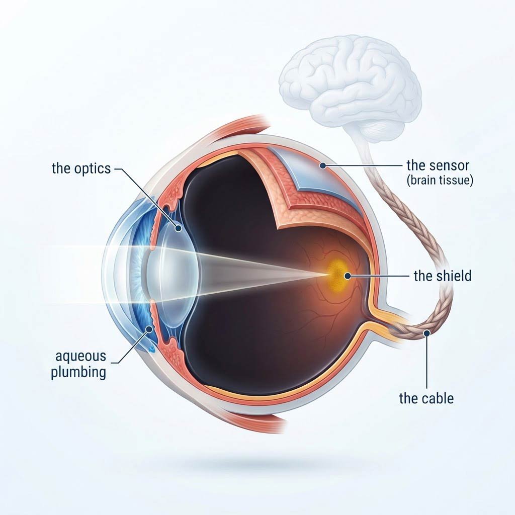
The architecture: the cornea (the fixed outer lens — two-thirds of the eye's focusing power) and the crystalline lens (the autofocus — shape-changed by ciliary muscles, stiffening lifelong into presbyopia), focusing light through the vitreous gel onto the retina — a ten-layer sheet of 126 million photoreceptors (120 million dim-light rods, 6 million color cones), supported by the retinal pigment epithelium (the gardener layer), with the macula's cone-dense fovea as the high-resolution center and the optic nerve exiting as a million-fiber cable toward the visual cortex. The front chamber runs its own plumbing: aqueous humor secreted by the ciliary body, drained through the trabecular meshwork — a balance that sets the intraocular pressure.
The Anatomy of the Camera and Its Brain
The optics are a two-lens system. The cornea does most of the focusing — a fixed, avascular, transparent window (its nourishment arrives via tears and aqueous, not blood — the engineering that keeps it clear) and the fastest-healing tissue in the body. The crystalline lens fine-tunes: a flexible, growing-all-your-life structure whose shape is adjusted by the ciliary muscles for near focus. Presbyopia — the reading-glasses sentence of middle age — is simply the lens stiffening with decades of accumulated layers, the autofocus losing its range. No disease, just arithmetic; the reason every eye over 45 needs help at arm's length.
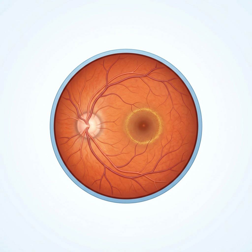
The sensor is where the eye stops being a camera and becomes an outpost of the central nervous system. The retina is a ten-layered processor — photoreceptors at the back, layers of wiring and computation above them — that performs more processing before a signal ever reaches the brain than a naive camera metaphor suggests. The fovea, at the macula's center, is pure cones packed at maximum density: a 0.35 mm island where the wiring is pushed aside and every photoreceptor gets a private line — the high-resolution zone you are using to read this sentence, moved word by word without your noticing. The rods own the periphery and the night: so sensitive that single-photon detection has been demonstrated in the dark-adapted eye, at the cost of sharpness (rods share wiring — many sensors, one line — which is why the night sky's dimmest stars vanish when looked at directly).
The support systems carry the organ's disease stories. The retinal pigment epithelium (RPE) is the gardener: it recycles photoreceptor outer segments (which are shed and rebuilt on a daily cycle — your light sensors are replaced throughout life), shuttles nutrients, and disposes of waste — its gradual age-related dysfunction is dry AMD's engine. The aqueous plumbing — production by the ciliary body, drainage through the trabecular meshwork — sets the intraocular pressure; when drainage fails, pressure rises silently against a nerve that does not complain, and glaucoma steals the visual field from the outside in (Chapter 2's story). And the optic nerve — a million fibers of CNS white matter — exits at the blind spot, the one patch each eye cannot see, papered over by the brain's interpolation. The eye is not a window with a viewer behind it; it is a brain-adjacent computer wearing a lens.
Photoreceptors
126 million sensors — rods for the night, cones for the fine print
RPE
The gardener layer — recycling sensors daily, failing quietly in AMD
The Macula
The high-res center — shielded by plant pigments the body cannot make
Optic Nerve
A million CNS fibers — the cable glaucoma silences
The Eye as the Body's Window — and Its Ledger
Four consequences of this architecture organize everything the rest of the page teaches:
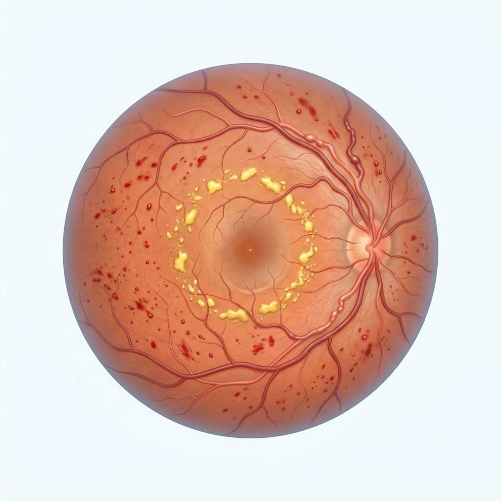
- The retinal exam sees the whole body: the eye is the only organ where microvasculature and nerve tissue are directly visible. Diabetes, hypertension, atherosclerosis, elevated intracranial pressure, and multiple sclerosis all write on the retina — the dilated exam is a systemic screening wearing the costume of a vision test.
- The diabetic ledger is silent until it is late: diabetic retinopathy — microaneurysms, leaks, exudates, and eventually new fragile vessel growth — accumulates for years without a single symptom, because the retina has no pain receptors and central vision survives until the macula is involved. This is why the annual dilated exam is non-negotiable in diabetes (Chapter 2), and why 'my vision is fine' is the sentence the exam exists to overrule.
- The macular shield is dietary, and the shield is real: lutein and zeaxanthin are the only carotenoids the body deliberately selects and concentrates in a tissue — filtering blue light and quenching photo-oxidative chemistry in the exact spot it does the most damage. The body cannot make them; the macula's yellow armor is built from the plate (kale, spinach, egg yolks — Chapter 3's arithmetic).
- The childhood-light story is the modern epidemic's mirror: time outdoors in childhood protects against myopia onset — the East Asian and trial data converge on roughly 2 hours daily of outdoor light slowing axial eye growth. Screens do not 'damage' the eye structurally (the honest note this page keeps), but the indoor life they anchor removes the protective exposure — the myopia generation's counterintuitive dose: light, not less reading.
Your retina is a piece of your brain you can look at — 126 million sensors, rebuilt nightly, armored with plant pigments it cannot make, wired by a million fibers to the cortex. Guard the macula, the glucose, and the pressure: the eye keeps score for the whole body.