Inside the joint.
Where does osteoarthritis actually happen in the body?
Osteoarthritis lives in the synovial joint — but the crucial anatomy lesson is a paradox: the cartilage that OA destroys has no blood vessels and no nerves. It feeds by cycling load — compression squeezes waste out, release draws nutrient-rich synovial fluid in. That is why the disease both causes and punishes inactivity: a resting joint starves its own cartilage, and pain that discourages movement quietly accelerates the process it signals.
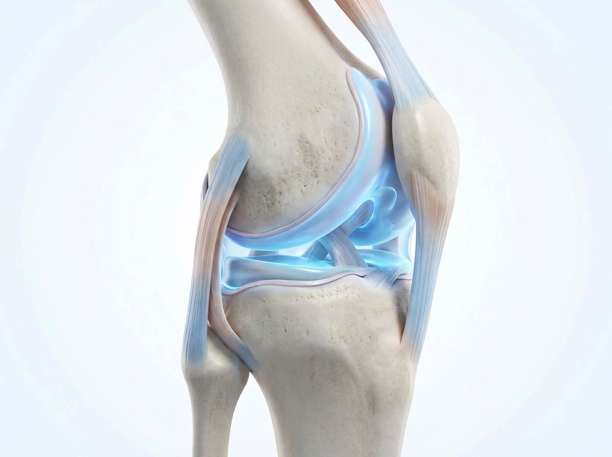
A sealed, self-lubricating system: bone ends capped with glassy cartilage, cushioned by the meniscus (in the knee), wrapped in a capsule lined by synovium — the membrane that manufactures the fluid everything inside depends on. No blood vessels cross into cartilage; the joint feeds itself through movement.
The Anatomy of a Self-Lubricating System
Articular cartilage is one of engineering's most improbable materials: a water-saturated matrix of collagen and proteoglycans, slicker than any man-made bearing, able to absorb decades of impact. But its design choice defines its vulnerability — it is avascular. Every nutrient, every repair material must arrive by diffusion from synovial fluid, and that diffusion is driven by load cycling: compression squeezes fluid out, relaxation draws fresh fluid in. A joint that moves is a joint that eats.
Beneath the cartilage sits subchondral bone — living bone that remodels under load and stiffens as OA progresses, transmitting more shock upward into the cartilage it was meant to cushion. Around it, the synovium — normally a quiet factory producing lubricant — becomes an inflammation source in OA, and the meniscus (the knee's C-shaped shock absorber) both cushions and, when torn, accelerates the breakdown. The four tissues fail together, because they load together.
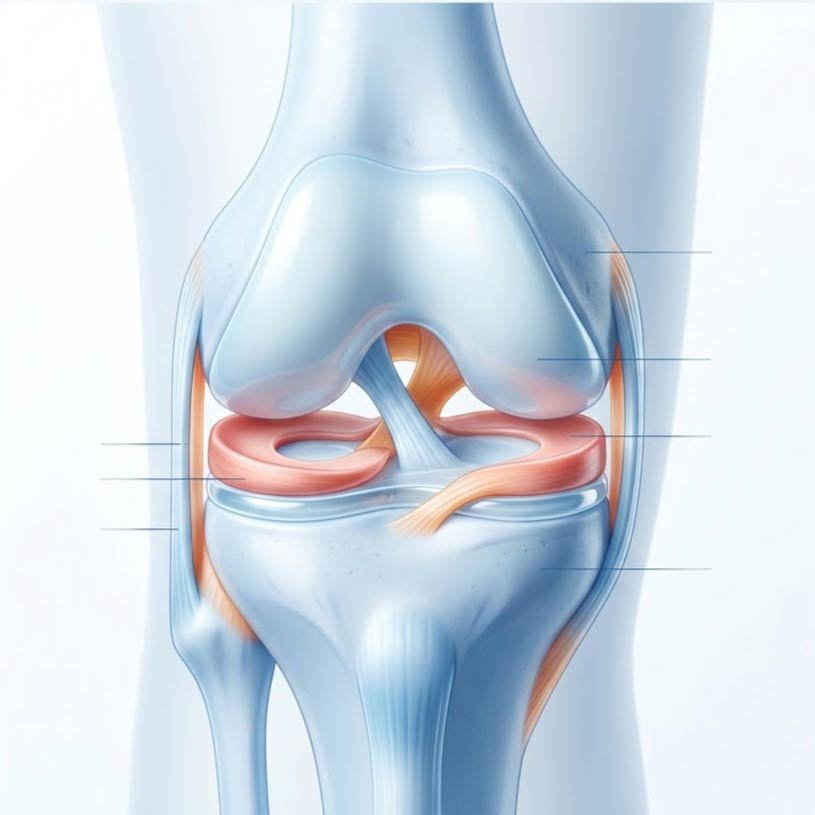
Cartilage
The glassy shock absorber — avascular, aneural, fed by motion
Subchondral Bone
The stiffening foundation beneath — remodels under load
Synovium
The fluid factory — and an inflammation source in OA
Muscles & Meniscus
The primary shock absorbers — the modifiable layer
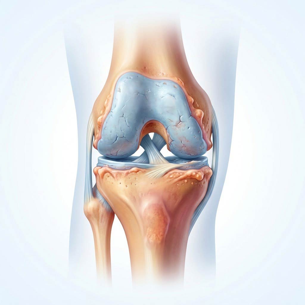
From Load to Breakdown: The Progression Sequence
The failure cascade involves every tissue in the joint, feeding back on itself:
- Matrix depletion: chondrocyte enzyme release tips cartilage from maintenance mode into degradation mode — the water content and resilience fall.
- Fissuring: the softened surface cracks under load, roughening the once-glassy bearing.
- Bone response: subchondral bone stiffens and develops cysts; the joint edges grow osteophytes — bony spurs that stabilize but limit motion.
- Synovial inflammation: breakdown products irritate the synovium, adding inflammatory mediators to a mechanical problem.
- Muscle withdrawal: pain inhibits the muscles that protect the joint — loads shift from muscle to cartilage, and the cycle accelerates. This last step is the most treatable one in the entire sequence.
Cartilage has no blood vessels and no nerves — it cannot bleed, cannot feel, and cannot feed itself except through movement. The pain of osteoarthritis comes from everything around the cartilage, and the cure for the cartilage is the very motion the pain discourages.