Inside the living reservoir.
What is the bladder — and why is the bag that holds the body's rejects not actually a bag at all?
The bladder is a muscular reservoir of remarkable engineering: a sac that stretches from golf ball to grapefruit (400–600 mL), lined with the most non-stick surface in the body (the urothelium — umbrella cells in a waterproof, stretchable GAG-coated mosaic), wrapped in a smooth-muscle pump (the detrusor), emptied through a double gate (one involuntary sphincter, one voluntary — the only autonomic function in the body with a manual override), and coordinated by a command center in the brainstem. It holds, sterile, a concentrate of everything the blood has discarded — the waste the kidneys filtered (our kidneys page's 180 liters distilled to 1–2) — and it does so while being one of the most infection-attacked surfaces in biology. This page teaches the organ; our UTI page teaches the war fought on it.
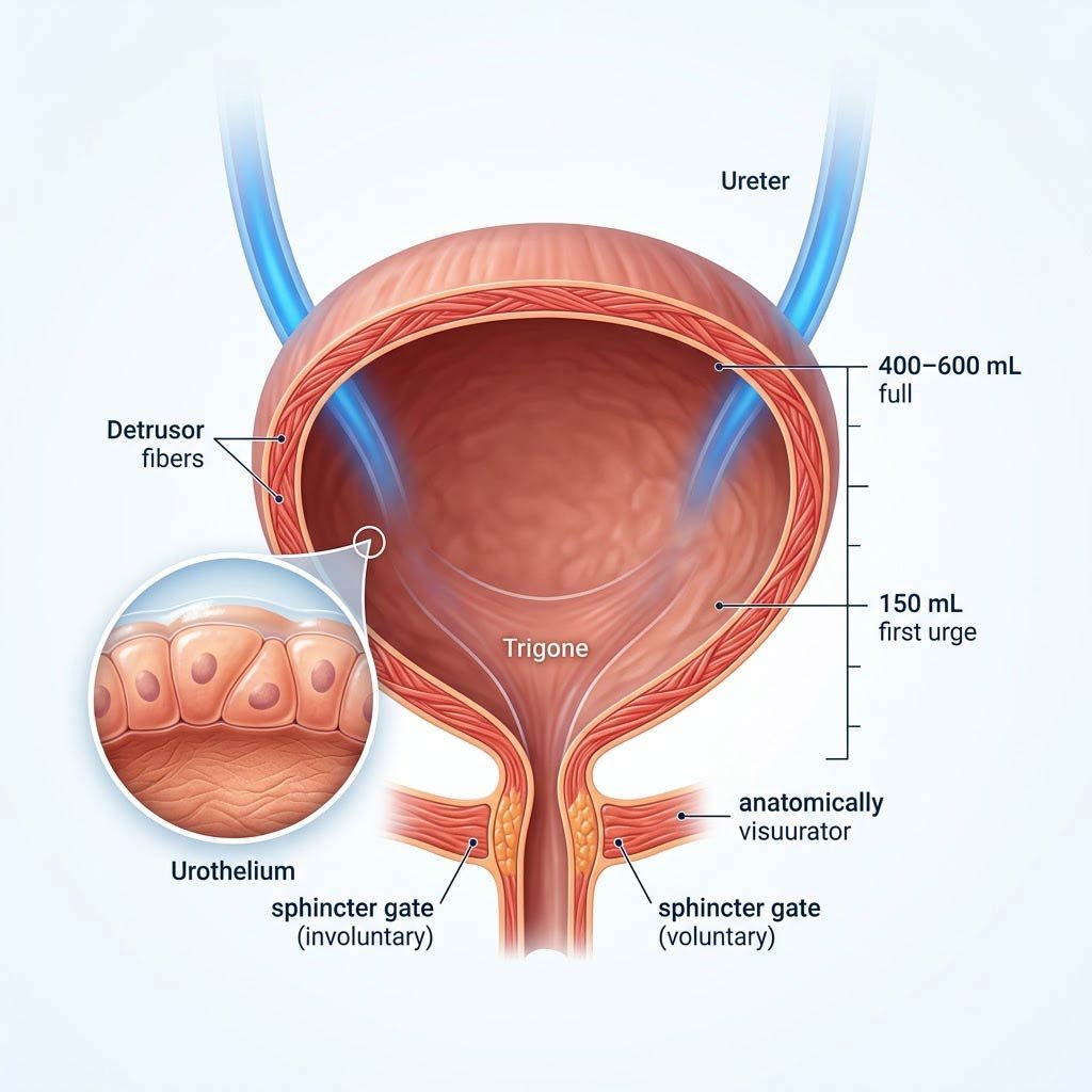
The architecture: a detrusor muscle shell (three interlacing smooth-muscle layers — the pump), a urothelial lining (umbrella cells with uroplakin tiles that unfold as the organ fills, sealed beneath a glycosaminoglycan non-stick coat), the trigone (the triangular base where both ureters enter and the urethra leaves), and the sphincter pair — internal (autonomic) and external (voluntary, skeletal muscle, trained like any muscle). Urine arrives drop by drop from the kidneys; the first urge registers around 150–300 mL; capacity is roughly 400–600 mL.
The Anatomy of Hold and Release
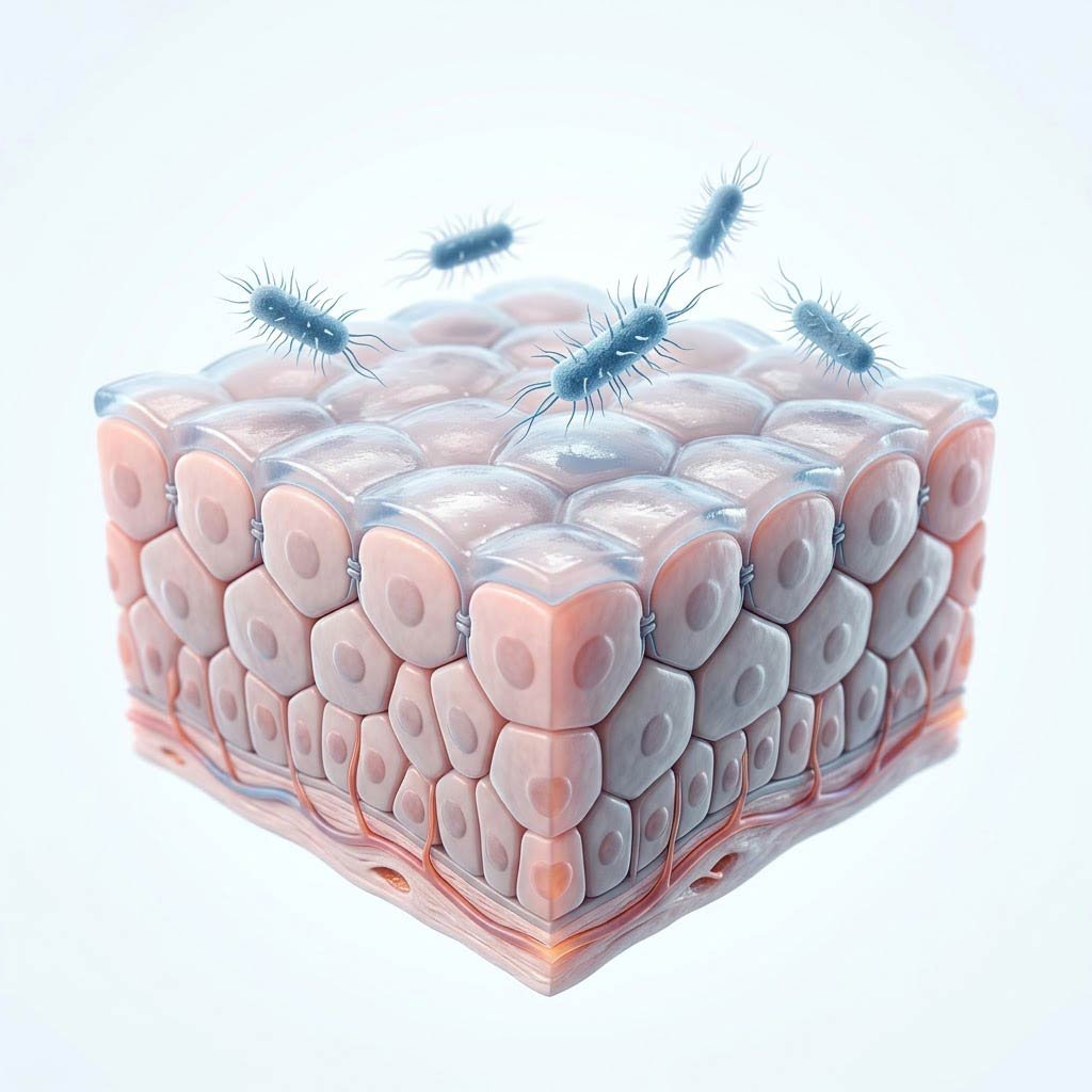
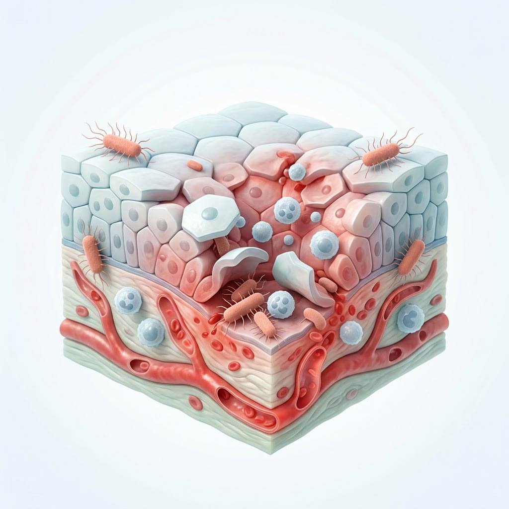
The lining is the organ's masterpiece. The urothelium is a transitional epithelium whose surface cells — umbrella cells — carry plaques of uroplakin protein arranged like waterproof tiles; as the bladder fills, these tiles unfold from folded reserves and the cells flatten, increasing surface area several-fold without losing their seal. Beneath and above runs the glycosaminoglycan (GAG) layer — the non-stick coat our UTI page teaches from the infection side: it holds urine's ammonia, urea, and variable osmolality away from the nerve-rich tissue below, and it denies bacteria the grips they need. The result is a biological paradox: a permeable-looking membrane that is among the tightest barriers in the body — blood on one side, toxic concentrate on the other, nothing crossing.
The machinery around it is equally deliberate. The detrusor — three interlaced layers of smooth muscle — stays relaxed and compliant through the fill (the bladder spends 97% of its day holding, not voiding), then contracts as one coordinated wave to empty. The dual sphincter at the exit: the internal sphincter runs on autopilot; the external sphincter is skeletal muscle, under voluntary command — the structure that makes continence a learned skill and its loss a treatable failure. The wiring runs to the pontine micturition center in the brainstem, which orchestrates the voiding reflex — and which descending pathways from the brain can veto or trigger on command. Infants run the reflex automatically; toilet training is, quite literally, the cortical override being installed.
Umbrella Cells
The stretch tiles — uroplakin mosaics unfolding as the organ fills
Detrusor
The pump — relaxed 97% of the day, one coordinated wave to empty
Stretch Receptors
The volume gauge — the first urge is data, not an emergency
The Sphincters
The dual gate — one autonomic, one voluntary and trainable
The Cycle, The Clock, and The Night
Three operational facts complete the organ's story — the ones every habit in Chapter 3 is built on:
- The flush is rate-dependent: urine that leaves every 3–4 hours keeps contact time short and concentration low; the held bladder gives bacteria a warm, standing pool — the reason 'don't hold it' is mechanics, not manners (and the same lesson our kidney-stones page teaches for stone chemistry).
- The night runs on a hormone: antidiuretic hormone (ADH) rises overnight, telling the kidneys to concentrate urine and reduce volume — the reason most people sleep 7–8 hours without voiding. Nocturia (waking twice or more to void) has many causes — prostate enlargement in men, fluid timing, sleep apnea (our sleep-apnea page's under-taught link), heart failure — and deserves the workup, not the shrug.
- The organ ages: capacity modestly shrinks, the voiding reflex grows more assertive, and in men the prostate — wrapping the urethra like a hand around a garden hose — enlarges with age (the men-over-40 conversation, our men-over-40 topic's engine). None of this is 'just aging' in the sense of untreatable; the treatment tier for every item is real.
- The floor beneath it matters as much as the wall: the pelvic floor — the muscular hammock holding bladder, uterus, and rectum — is the organ's shelf. Its weakness (childbirth, age, chronic straining) is incontinence's most common mechanism; its training is the most evidence-backed fix on this page (Chapter 3's Kegel story).
Your bladder holds half a liter of the blood's rejects behind the most non-stick wall in the body — and empties on command through the only autonomic circuit with a manual override. The reservoir is alive: guard the wall, train the floor, respect the flush.