Inside the collecting system.
Where do stones actually happen in the body?
A stone's biography runs through the renal collecting system: the calyces (where crystals form and grow, sometimes for years, painlessly), the renal pelvis (the funnel they must enter), and the ureter — a 25-cm muscular tube with three natural narrowings, each a potential choke point. The kidneys themselves feel no stone until it moves; the ureter feels everything. Understanding the geography explains the pain's location, its radiation, and why 'it has to pass' is a sentence with three checkpoints.
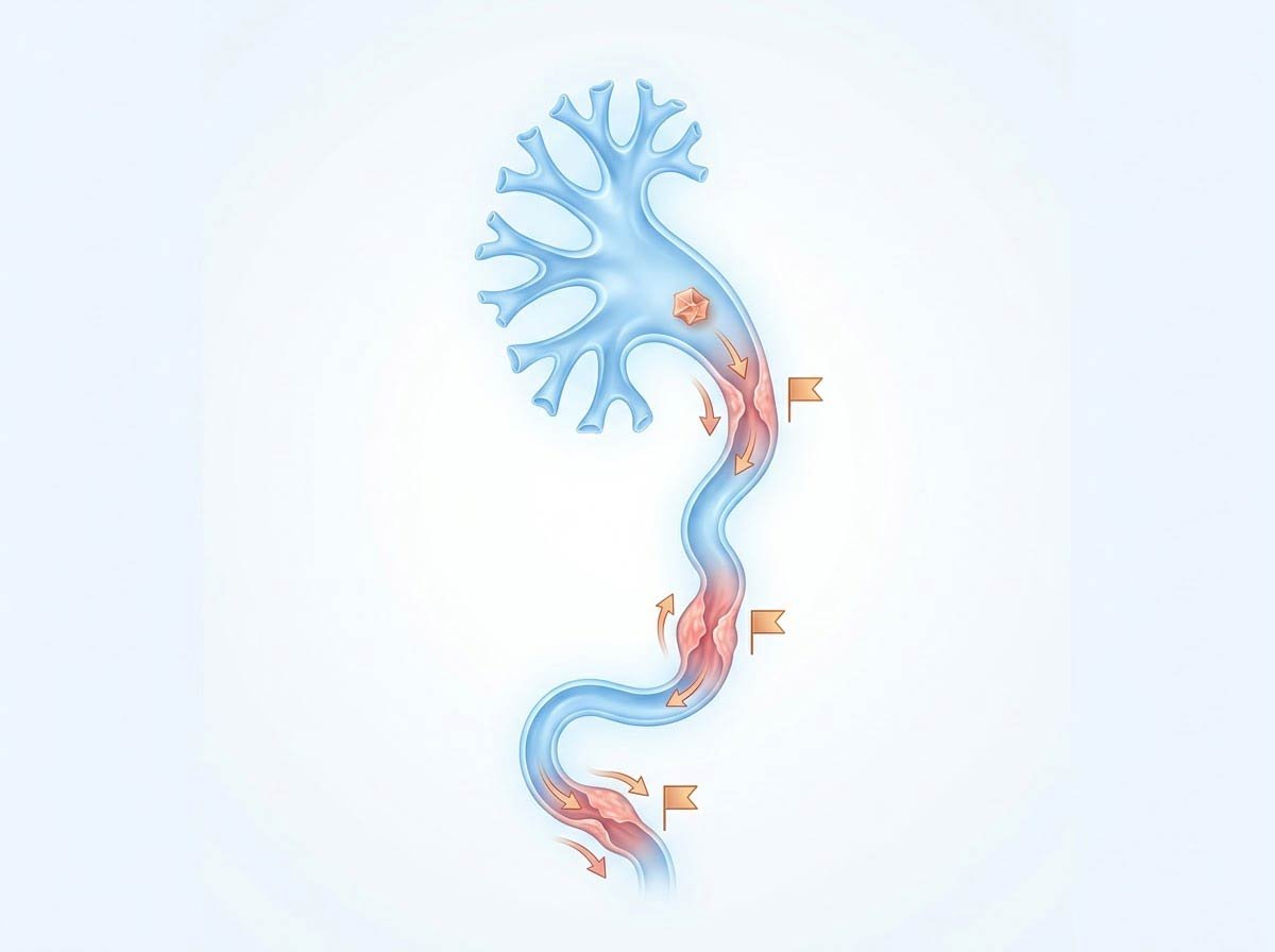
The kidney filters ~180 liters of plasma daily, fine-tunes it to 1–2 liters of urine, and delivers it through a calyceal system into the ureter. Concentration is the system's design job — and supersaturation is its failure mode. The ureter's three narrowings (ureteropelvic junction, pelvic brim crossing, ureterovesical junction) are the passage's rapids.
The Anatomy of Concentration
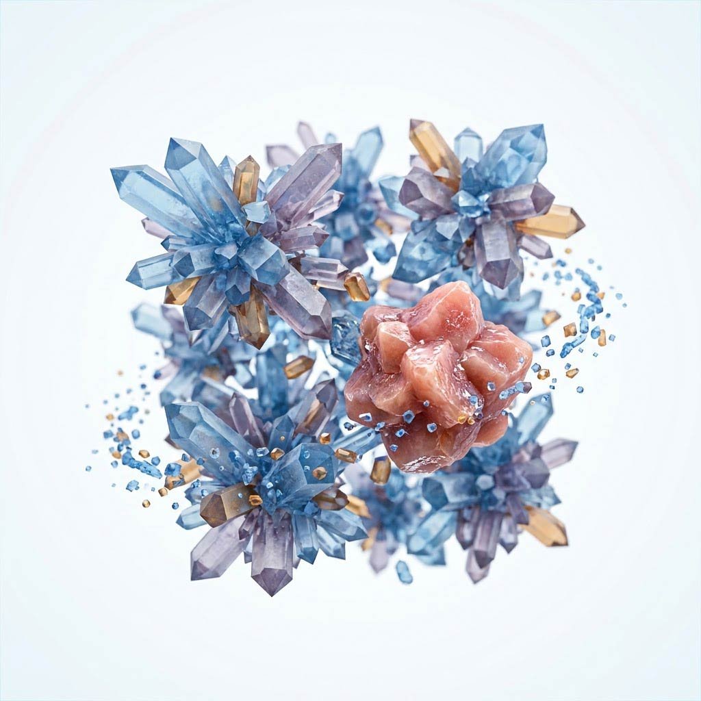
The kidney's concentrating machinery is its genius and its vulnerability: the loop of Henle builds a concentration gradient that lets the body reclaim water — and the same gradient concentrates stone minerals in the collecting ducts. In the renal papillae, where urine is most concentrated, calcium deposits (Randall's plaques) form and serve as stone templates — visible on analysis as the nidi of most calcium-oxalate stones. Dehydration doesn't just lower urine volume; it drives the concentrating system harder at exactly the tissue interface where stones start.
The ureter is the passage's gauntlet. A muscular, peristaltic tube ~25 cm long, it narrows at three points: where it leaves the kidney (ureteropelvic junction), where it crosses the pelvic brim (the iliac crossing), and where it enters the bladder (the ureterovesical junction — the tightest). A stone ≤5 mm usually passes; larger stones lodge, and the ureter's response is spasm — smooth muscle contracting against a rigid obstruction, the mechanism of renal colic: the severe, wave-like flank pain radiating to the groin that sends people to emergency departments writhing. The kidney itself: silent. The ureter: everything.
Nephrons
The 180-liter filter — and the concentration gradient that concentrates stones
Renal Papillae
Randall's plaques — the stone templates at the concentration peak
Ureter
The 25-cm gauntlet — three narrowings, one spasm response
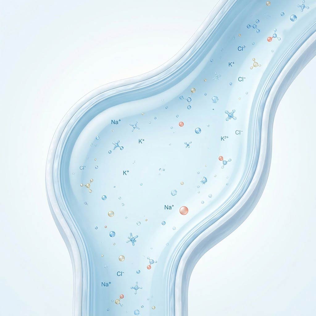
Citrate
The built-in inhibitor — binds calcium, keeps crystals dissolved
From Crystal to Colic: The Event Sequence
A stone event assembles in stages, each with its own management:
- Formation: months to years of quiet crystal growth on papillary plaques — silent, preventable, invisible without the chemistry that predicts it.
- Dislodgement: the stone enters the funnel — first twinge, often mistaken for back strain until the pattern declares.
- Obstruction & colic: the ureter spasms against the blockade — the wave-like flank-to-groin pain, nausea, the restlessness that distinguishes colic from peritonitis (stone patients pace; perforated-belly patients lie still).
- Impaction or passage: ≤5 mm usually passes over days to weeks; ≥6–7 mm usually needs intervention — the size thresholds that guide expectant management versus lithotripsy, stenting, or ureteroscopy.
- The aftermath: passage is not cure — the chemistry that built the stone remains, which is why the 24-hour urine follows the strainer.
Your kidneys filter 180 liters a day and concentrate it to two — a design brilliant for survival and generous to stones. The entire prevention of stone disease is, at its core, a plea to that design: carry more water, and the chemistry never gets its chance.