Inside the living hinge.
What is a joint — and why did evolution build a bearing slicker than ice on ice, then forget to give it a blood supply?
A synovial joint is the most elegant machine in the body: two bone ends capped in hyaline cartilage — a bearing surface with a friction coefficient as low as 0.001, up to 100 times slicker than ice gliding on ice — bathed in synovial fluid (the oil), wrapped in a synovium-lined capsule (the factory that makes the oil and staffs the immune border), and held true by ligaments (the tension cables). But the design carries one fateful economy: cartilage has no blood vessels and no nerves. Its chondrocytes — less than 5% of its own volume — are fed entirely by cyclic compression pumping nutrient fluid through the matrix. This is the page's central teaching: the bearing feeds on movement. Motion is lotion, load is nutrition, and stillness is the slow rust. The joints host this library's two great condition stories — osteoarthritis (our osteoarthritis page) and gout (our gout page) — and both are, at bottom, failures of this machine's quiet maintenance.
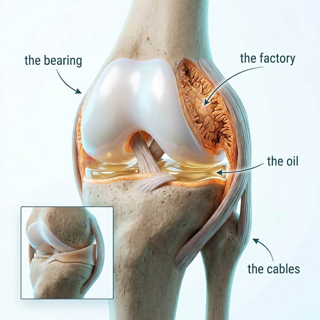
The architecture: hyaline cartilage (the glassy bearing — collagen II lattice swollen with water-holding proteoglycans, slicker than any man-made surface at joint speeds), the synovial membrane (a thin factory-and-immune organ whose cells secrete hyaluronic acid and lubricin into the joint space), synovial fluid (the viscous oil that lubricates and feeds the avascular cartilage), and the capsule and ligament cables that hold the geometry true under load. Knees add menisci — fibrocartilage shock absorbers that also distribute load.
The Anatomy of the Bearing and Its Economy
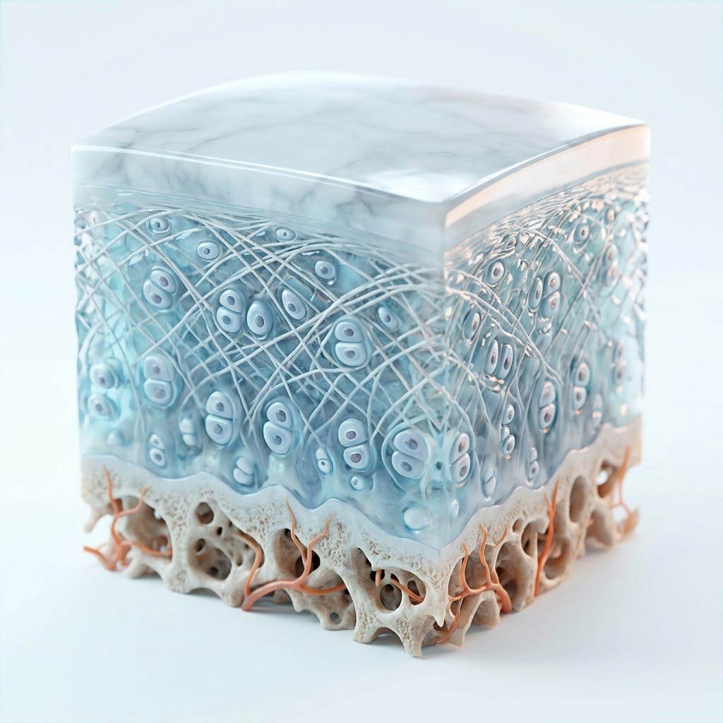
Cartilage is a composite material that engineers still cannot replicate: a collagen II lattice (the rebar) trapping proteoglycans — giant molecules whose negative charges pull water in like a molecular sponge (the hydraulics). Load the joint and the matrix compresses, expressing fluid; release it and the matrix re-expands, drawing fresh, nutrient-rich synovial fluid in. That cycle — compression, release, compression, release — is the cartilage's entire supply line. There is no capillary within it, no nerve ending on its surface, and its resident chondrocytes sit sparse and isolated, maintaining a matrix that turns over on the timescale of years, not days. The consequences organize this whole page: the bearing cannot inflame (no nerves), cannot bleed, cannot mount a fast repair — and it starves in stillness. The man on the couch is not resting his knees; he is unplugging them.
Around the bearing, the synovium runs the joint's factory and border control: its cells manufacture hyaluronic acid (the fluid's viscosity) and lubricin (the boundary lubricant), while its macrophage sentinels survey the space for crystal debris and cartilage fragments. It is also the battlefield of the inflammatory arthritides — in rheumatoid arthritis, the immune system attacks this lining, and the factory becomes a destruction crew (Chapter 2's story). The menisci of the knee — the wedges of fibrocartilage that distribute load — carry the same avascular economy in miniature: their outer rim has blood supply (the 'red zone' that can heal), their inner two-thirds do not (the 'white zone' that cannot — why meniscus tears are managed so differently by location).
And the machine has one more economy that completes the story: the surfaces are congruent by design but not by accident — ligaments hold them aligned, and load passing through the aligned joint is distributed across the whole bearing. Injure the cable (an ACL tear), or chronically overload one edge of the bearing (obesity, malalignment), and the same total load concentrates on a fraction of the surface — the pressure arithmetic behind post-traumatic and weight-driven osteoarthritis. The joint is a precision instrument: it forgives enormous load through alignment and motion, and punishes modest load through misalignment and stillness.
Hyaline Cartilage
The bearing — slicker than ice, fed by compression, not blood
Synovium
The factory floor — lubricant production and immune border in one
Synovial Fluid
The oil — hyaluronic acid soup that feeds the avascular bearing
Capsule & Ligaments
The tension cables — holding the hinge true under load
Beyond the Hinge: The Rules the Rest of the Page Runs On
Four consequences of the bearing's economy explain everything from symptoms to prevention:
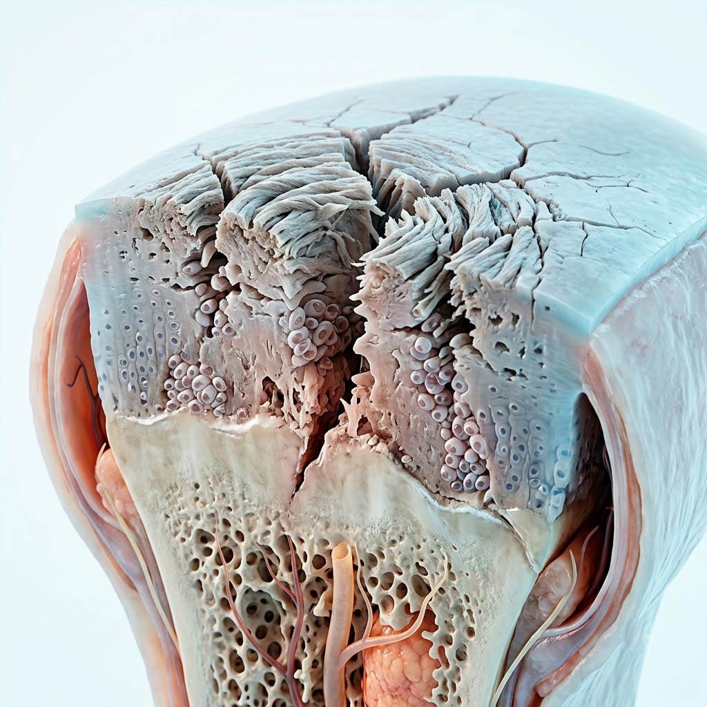
- No nerves in cartilage — the disease is silent until it isn't: OA damages the bearing painlessly for years; pain arrives when the process reaches the bone and synovium beneath — where the nerves live. The X-ray never matches the timeline: by the time it hurts, the wear has history.
- The load arithmetic is brutal and modifiable: level walking loads the knee at 2–3× body weight; stairs and squats 3–4×. Every kilogram of body weight is 3–4 kilograms across the patellofemoral bearing at the stairwell — the arithmetic that makes weight the #1 modifiable OA variable (Chapter 3's Framingham story).
- The exercise paradox, resolved: recreational runners do NOT develop more knee OA than sedentary people — the cohort data and meta-analyses show less, not more (Chapter 3's case study). Cyclic, aligned, graduated load feeds the bearing; sudden unprepared load (the weekend-warrior pattern our muscles page retires) and chronic overload (obesity) damage it. 'Wear' was always the wrong metaphor — cartilage is not a tire; it is a gland that secretes health when compressed.
- The joint ages on two clocks: the intrinsic clock (chondrocyte senescence, water content falling, matrix turnover slowing) and the extrinsic clock (load history, injury, inflammation — the one you influence). Sarcopenia compounds both: the muscles that should decelerate forces across the joint (our muscles page's shock-absorber role) fade exactly as the bearing needs them most — the joint-muscle partnership aging together.
Your joints run on bearings up to a hundred times slicker than ice on ice — built without a single blood vessel, fed only by the pumping of movement itself. Motion is lotion. Load is nutrition. Stillness is the slow rust.